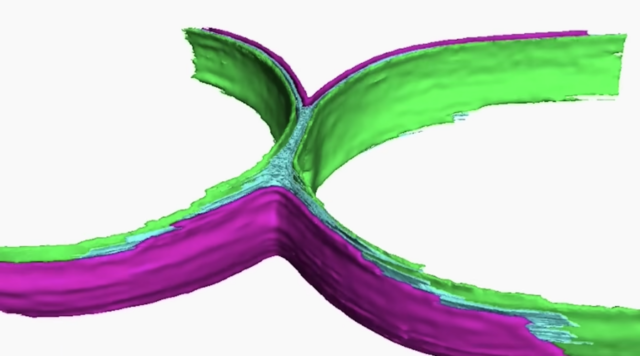
Look closely at the double-layered membranes of E. coli thanks to live cell fluorescent imaging and cryo-electron tomography. These revealing views of cell division, as well as up-close footage of the mutants that alter E. coli’s DNA, have never been seen before.
The images were captured by researchers in the labs of Luke Chao and Tom Bernhardt in the Blavatnik Institute at Harvard Medical School.

The work offers new insights into the division process and may aid in the fight against antibiotic resistance, since these drugs typically target bacteria as they divide, when the cell wall and membranes are weakest.
“Led by postdoctoral research fellows Paula Navarro and Andrea Vettiger, the two groups made the discoveries possible by combining their expertise in bacterial cell division, bacterial genetics, and cutting-edge imaging.”
Watch these related bacteria videos on TKSST:
• Bacteria Growth, a time lapse
• Subvisual Subway, the Art of NYC’s Bacterial World
• The Foldscope – A Paper Microscope that Costs $1
• Evolutionary branching in action: Bacteria adapt to antibiotics
Plus: Cell division in a frog egg.
Curated, kid-friendly, independently-published. Support this mission by becoming a sustaining member today.